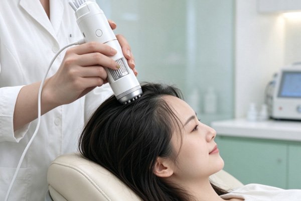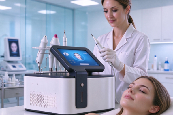
A skin analyzer uses image capture and data analysis to help assess the distribution, color, and appearance of facial pigmentation spots. Some devices also feature cross-polarized light or ultraviolet (UV) imaging to reveal skin pigmentation characteristics from different perspectives. For beauty clinics and equipment buyers, understanding the relevant indicators and imaging methods helps with interpreting reports, evaluating device capabilities, and supporting daily skin management.

Key Data Used by Skin Analyzers to Detect Pigmentation Spots
Pigmentation analysis mainly focuses on the spatial distribution and visual characteristics of pigmented areas. The specific data available depends on the device’s imaging configuration and analytical functions.
Pigmentation Area and Distribution
Area and location data help practitioners record basic information about pigmented areas on the face.
- Pigmentation area: Measures the extent of pigmented regions identified in an image to estimate their coverage.
- Distribution location: Shows whether pigmented areas are concentrated on the forehead, cheekbones, cheeks, or other facial regions.
- Number of regions: Records the distinct pigmented areas identified in an image to help assess their distribution patterns.
These data are useful for establishing baseline records, but they cannot independently determine the type of pigmentation.
Color and Contrast
Color characteristics help the device identify visual differences between pigmented areas and the surrounding skin.
- Color differences: Analyzes variations in hue between pigmented regions and nearby skin.
- Brightness: Compares the brightness of local areas to help identify pigmentation boundaries.
- Color parameters: Some devices use parameters such as RGB and L*a*b* to analyze image colors and may generate pigmentation-related scores.
Color parameters can help quantify image differences, but they do not directly represent the absolute amount of melanin inside the skin.
How Different Imaging Modes Help Identify Pigmentation Spots
Different light sources and imaging methods reveal different skin characteristics. Understanding the modes supported by a device helps practitioners interpret the scope and limitations of its results.
White Light and Cross-Polarized Light
White light is mainly used to observe the overall appearance of the face, while cross-polarized light can reduce some surface reflections and help reveal details in pigmented areas.
- White-light imaging: Shows the color, location, shape, and surrounding skin appearance of pigmentation spots.
- Cross-polarized imaging: Reduces some surface specular reflections to help observe pigmentation distribution and boundary characteristics.
- Image comparison: Comparing images captured in different modes provides information that may not be available from a single image.
Cross-polarized light can improve the visibility of certain features, but it cannot independently determine the tissue depth of pigmentation.
UV Imaging and Potential Pigmentation Features
Some skin analyzers support UV imaging, which can reveal pigmentation contrasts that differ from those seen under ordinary white light. Certain pigmentation features that are difficult to notice with the naked eye may appear more prominent under UV illumination. Combining UV images with other imaging modes can help assess facial pigmentation distribution. However, a UV image cannot directly prove that a pigmentation spot is located in deeper skin layers. Interpretation still requires consideration of the device’s capabilities and other available findings.
How to Interpret Pigmentation Analysis Reports Correctly
Images, area measurements, and scores in a detection report represent different types of information. Understanding their definitions and limitations helps prevent misinterpretation.
Distinguishing Image Data from Pigmentation Indicators
Devices may generate results through image recognition, color calculations, or optical analysis. Area, number, and location describe the pigmented regions identified by the system. Some devices calculate indices based on color differences or optical signals, with their specific meanings defined in the device documentation. Algorithms and scoring standards vary between devices, so their numerical results should not be compared directly across different systems. Beauty-oriented skin analysis results are intended to support observation and cannot replace professional medical evaluation.
Tracking Changes Through Repeated Measurements
Regular measurements help document changes in the appearance of pigmentation spots, but testing conditions should remain as consistent as possible for meaningful comparisons.
- Save historical records: Keep testing dates, images, and relevant indicators to track changes over time.
- Standardize imaging conditions: Maintain consistent light modes, device settings, shooting distance, and facial positioning whenever possible.
- Compare results comprehensively: Review images and data from different dates to observe changes in pigmented areas.
- Interpret differences cautiously: Lighting, skin condition, and image-recognition algorithms can affect results. A change in a single measurement does not necessarily indicate improvement or worsening.
Consistent recordkeeping improves the comparability of results and provides useful information for routine skin management.
How to Choose a Skin Analysis Device for Pigmentation Assessment
When purchasing equipment, beauty clinics should consider their service requirements and evaluate imaging configurations, analytical functions, report presentation, and data management capabilities.
Evaluate Imaging Modes and Analytical Functions
A device’s light-source configuration affects the image information it can capture. Buyers should verify the actual functions available before making a decision.
- Confirm light-source options: Check whether the device supports white light, cross-polarized light, UV light, or other imaging modes.
- Review analytical indicators: Check whether it provides pigmentation area, distribution, color characteristics, and relevant scores.
- Understand data definitions: Confirm how indicators are calculated and what they are designed to measure. Avoid interpreting image-based scores as absolute melanin content.
- Examine reporting functions: Evaluate image annotations, data explanations, and historical comparison features against the clinic’s needs.
Selecting imaging modes and analytical functions that match actual business requirements helps maximize the practical value of the equipment.
Consider Workflow Efficiency and Routine Maintenance
Operating efficiency and maintenance requirements also affect the long-term user experience.
- Operational efficiency: Review the image capture, mode-switching, and report-generation processes to determine whether they fit the clinic’s workflow.
- Data management: Check whether the device supports customer image storage, historical record retrieval, and periodic comparisons based on business needs.
- Maintenance requirements: Confirm the specific requirements for cleaning, calibration, and routine maintenance.
- Technical support: Review the availability of product training, troubleshooting, and after-sales services.
A comprehensive evaluation of analytical functions, ease of operation, and maintenance requirements helps clinics select equipment suited to their needs.
When detecting pigmentation spots, skin analyzers primarily assess area, distribution, color, and related pigmentation indicators. Some devices also use cross-polarized light and UV imaging to provide additional visual information. Different imaging modes reveal different optical characteristics and cannot independently determine pigmentation depth or diagnose a specific skin condition. Beauty clinics should evaluate imaging configurations, reporting functions, and data management capabilities according to their actual requirements. Guangzhou Cosnext Technology Co.;Ltd provides product and technical solution consultations for beauty equipment buyers, helping them explore suitable configurations for pigmentation analysis.