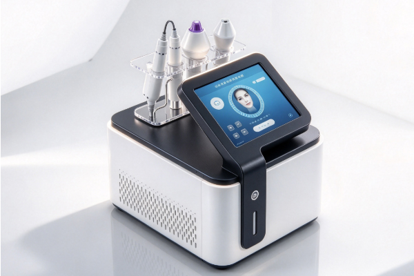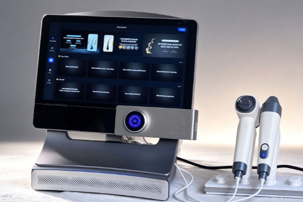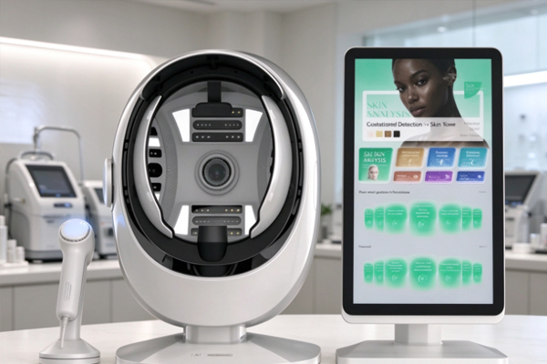
Skin analyzers use facial imaging and image analysis to display selected skin surface features, helping beauty clinics understand clients’ skin conditions. Report contents vary depending on the device’s light sources, imaging methods, and software functions. Common items include facial images, pore appearance, wrinkle and texture features, pigmentation distribution, and areas of redness. Understanding these indicators and their intended uses helps users interpret results appropriately and supports client communication, skincare planning, and follow-up comparisons.
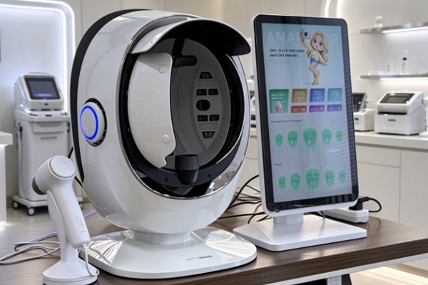
Basic Information in a Skin Analyzer Report
Reports typically record client information and facial images to document each assessment and support future reviews and comparisons.
Client Information and Assessment Records
Basic information helps organize client records and makes it easier to retrieve assessments from different dates.
- Client Information: Depending on the device and recordkeeping needs, reports may include a client ID or other necessary details to distinguish individual records.
- Assessment Date: Recording the date provides a time reference for observing changes in skin appearance.
- Assessment Settings: Some devices record imaging modes, light source types, or related parameters to help clarify the conditions under which images were captured.
Client information should be stored securely, with access controlled according to the clinic’s data management requirements.
Facial Images and Imaging Modes
Facial images are an important part of the report. Different imaging methods can reveal different optical characteristics of the skin.
- Standard Images: Show areas such as the forehead, cheeks, nose, and chin, making it easier to observe visible spots, pores, and skin texture.
- Polarized Images: Different polarization methods alter the presentation of surface reflections, helping users observe skin surface features and certain pigmentation- and redness-related characteristics.
- Specific Wavelength Images: Some devices use ultraviolet light or other specific wavelengths to reveal fluorescence or other optical differences, providing additional information for observing certain skin features.
Images captured using different modes are not directly interchangeable. Interpretation should be based on the device’s imaging principles and stated functions.
Key Indicators in a Skin Analysis Report
Reports may convert selected skin features into numerical values, grades, or marked areas. The meaning of each result depends on the analysis method used by the device.
Pores, Wrinkles, and Skin Shine
These indicators are mainly used to assess the visible appearance of the skin surface.
- Pore Appearance: Some devices analyze the number, area, or distribution of visible pores. Results may be affected by image clarity and recognition algorithms.
- Wrinkles and Texture: Systems may use image contours, contrast, and texture features to display fine lines, wrinkles, or surface roughness.
- Skin Shine: Devices can analyze the distribution of surface shine based on image reflections, but this should not be treated as a direct measurement of sebum production.
These indicators help users observe changes in skin appearance but cannot replace direct measurements or professional assessment.
Pigmentation, Redness, and Fluorescence Features
Some devices use specific light sources and image analysis algorithms to display color and fluorescence differences, helping users identify areas that may warrant closer observation.
- Spots and Pigmentation: Reports may show the distribution, number, or area of visible spots. Certain imaging modes may also assist in analyzing pigmentation-related features.
- Redness: Systems can mark areas of facial redness to show their distribution, but images alone cannot determine a specific medical condition.
- Fluorescence Features: Some devices display image features associated with fluorescent substances such as porphyrins. These results may be affected by imaging conditions and other factors.
These findings are intended to support observation. They should not be used alone to infer skin depth, bacterial counts, or medical diagnoses.
How Do Skin Analyzers Present Results
In addition to original images, some devices provide scores, marked regions, and historical comparisons to help users review the results.
Individual Scores and Overall Summaries
Scoring functions can present selected analysis results in a consolidated format, making individual skin features easier to review.
- Individual Scores: Devices may assign scores or grades to indicators such as pores, wrinkles, or pigmentation. Their meanings depend on the device’s definitions.
- Overall Summaries: Some reports combine multiple analysis items to help users review the main findings at a glance.
- Evaluation Rules: Scores may be generated through image recognition, feature calculations, or preset evaluation rules. Not all indicators represent directly measured physiological data.
Algorithms and evaluation standards can vary between devices, so scores should not be compared directly across different systems.
Marked Regions and Historical Comparisons
These functions help users identify areas of interest and review changes over time. Region markers use colors, outlines, or graphics to indicate skin features. Devices with historical record functions can display images from different assessment dates side by side, making changes in appearance easier to observe. Some systems also organize historical data to show how selected indicators change over time.For more meaningful comparisons, keep cleansing conditions, facial expressions, imaging modes, and device settings as consistent as possible, while minimizing the effects of residual makeup or sunscreen.
How to Evaluate the Practical Value of a Skin Analysis Report
A report’s usefulness depends on the clarity of its indicator definitions, the consistency of the assessment process, and its suitability for routine management needs.
Check Imaging Principles and Indicator Definitions
Understanding where the data comes from and what it represents can help prevent misinterpretation.
- Understand the Imaging Methods: Confirm the device’s light sources, polarization modes, and specific wavelength functions. Distinguish original images from algorithm-based analysis results.
- Review Indicator Definitions: Check the analysis basis for pores, pigmentation, redness, and skin shine to avoid treating different types of data as equivalent.
- Understand the Limits of the Results: Image analysis cannot directly replace quantitative sebum measurement, lesion-depth measurement, or medical diagnosis. If a diagnosis is needed, assessment should be performed by an appropriately qualified professional.
Clear indicator definitions support the appropriate use of report information.
Evaluate Assessment Procedures and Data Management
Consistent preparation and organized recordkeeping help improve the reference value of reports. Follow the device’s instructions and keep cleansing conditions, skin appearance, and product residue as consistent as possible. Check whether the device can save assessment results and retrieve historical records according to the clinic’s needs. Images, indicator explanations, and data displays should be clear and easy to understand, supporting client communication and record management.
Skin analyzer reports typically include basic client information, facial images, pore and texture features, pigmentation and redness indicators, scores, and historical comparisons. The actual contents depend on the device configuration. When interpreting results, distinguish image presentation from algorithm-based analysis and keep assessment conditions consistent to make follow-up comparisons more meaningful. Guangzhou Cosnext Technology Co.;Ltd provides information and selection support for beauty equipment, helping clinics understand device functions according to their needs.