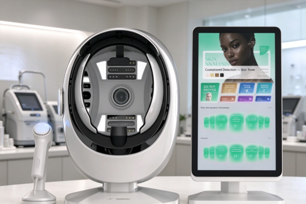
Hair analyzers are commonly used in scalp care, hair condition observation, and professional care consultations. They use magnified imaging to observe visible features such as the scalp surface, follicular openings, hair condition, and oil distribution. During scalp cleansing, oil-control treatments, moisturizing care, or other scalp care procedures, comparing images taken before and after treatment can help staff understand changes in the scalp surface and provide a reference for subsequent care. However, hair analyzer results can be affected by factors such as hair cleanliness, scalp moisture, ambient lighting, lens position, and operating methods.
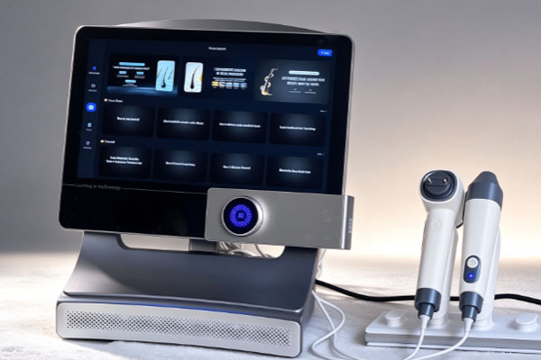
If different imaging conditions are used before and after treatment, even noticeable changes in the images do not necessarily indicate corresponding changes in the scalp itself. Therefore, when using a hair analyzer, it is important to pay attention to preparation before testing, the timing of post-treatment imaging, device cleaning, and image recording methods to keep testing conditions as consistent as possible. For beauty salons, scalp care centers, and professional hair analysis facilities, standardized use of a hair analyzer can improve the accuracy of image observation and make before-and-after comparisons clearer. Choosing equipment with stable imaging, clear magnification, and convenient image storage can help staff record visible scalp changes and provide clients with a more intuitive explanation of their care results.
Prepare Properly Before Scalp Care
Maintain the Original Scalp Condition
Before scalp care, it is recommended to use a hair analyzer to capture initial images and record the scalp condition before treatment begins. Avoid washing the scalp or applying new care products immediately before testing, as these actions may alter surface oil, moisture, and residue levels, reducing the value of the initial record as a reference.
Review recent care routines: Ask clients when they last washed their hair and whether they have used hair oil, scalp serums, styling products, or other care products.
Choose suitable testing locations: Depending on the care requirements, examine areas such as the hairline, crown, and scalp parting, and record the exact locations.
Reduce external interference: Before imaging, move hair away from the testing area to prevent strands, product residue, or visible debris from interfering with observation.
If scalp oil levels need to be compared before and after treatment, record the interval since the last shampoo and any products used. Differences in scalp cleanliness can directly change how oil appears in images, so a single scan should not be used to determine a fixed scalp condition.
Check the Device and Imaging Conditions
The cleanliness of the lens, lighting intensity, and magnification level of a hair analyzer can all affect image quality. Before testing, check that the device is functioning properly and adjust its settings according to the manufacturer’s instructions.
Clean the probe: Use cleaning products suitable for the device materials to clean contact surfaces and prevent residue from obstructing the lens.
Standardize imaging parameters: Set the magnification level, lighting mode, and image clarity settings according to the testing requirements to facilitate before-and-after comparisons.
Record images properly: Save the original images and label them with the testing date, observation location, and any necessary scalp care information.
For clients who require repeated testing, it is recommended to maintain separate image records for each individual. Consistent imaging locations and recording methods can reduce errors during follow-up tests and help staff explain directly observable changes to clients.
Pay Attention to Imaging Time and Operating Methods After Scalp Care
Determine Testing Time According to the Treatment
After treatment, a hair analyzer should not automatically be used immediately for comparison. Different scalp care procedures may leave moisture, cleansing products, or care products on the scalp surface, which can change how images appear. For example, scalp cleansing may reduce some oil and surface residue; moisturizing products may create a shiny or moist appearance; and scalp serums may produce different reflections due to product residue in specific areas. These changes do not necessarily indicate lasting changes in the scalp itself.
The testing time can be selected according to the purpose of the treatment:
Cleansing treatments: If the goal is to observe surface conditions before and after cleansing, record the imaging time after treatment and try to keep the scalp dryness level consistent.
Moisturizing treatments: If products remain on the scalp surface, record their use to avoid interpreting product-induced shine as a change in the scalp’s natural condition.
Other treatments: Determine the testing time according to the characteristics of the procedure, device instructions, and professional operating standards. When necessary, save immediate post-treatment images separately from images taken during subsequent follow-up visits.
Post-treatment images are mainly used to observe visible surface changes. They should not be used alone to assess hair growth, follicular function, or the long-term effectiveness of scalp care.
Avoid Result Distortion Caused by Operating Differences
Post-treatment testing should follow the same operating procedures used before treatment as closely as possible. If the observation location, magnification level, or lighting mode changes, the appearance of follicular openings, flakes, and oil may also change, potentially affecting the assessment. During testing, keep the probe steady and avoid applying excessive pressure to the scalp. Areas with obvious swelling, redness, broken skin, pain, or other discomfort should not be subjected to unnecessary contact. Whether testing can continue should be determined according to the device instructions and professional operating standards.
Images taken before and after treatment should be viewed side by side, with particular attention to visible features in the same locations, such as surface residue, oil appearance, and the condition around individual hair strands. Staff should describe image differences objectively and should not conclude that a treatment has achieved its intended results based solely on local color changes or a single magnified image.
Prioritize Hygiene and Image Management During Testing
Hair analyzers are generally used close to the scalp, and the probes of some devices come into direct contact with the skin. Therefore, hygiene management and image recording are essential parts of the testing process. Standardized procedures can reduce the risk of cross-contamination and help maintain consistent testing records.
Clean the Probe and Protect the Skin
Before and after each client uses the device, clean and disinfect the probe and related contact surfaces according to the manufacturer’s instructions. Do not apply disinfectants that are unsuitable for the lens, coatings, or electronic components, as they may damage the equipment. For probes that come into direct contact with the scalp, confirm that their cleanliness meets the device requirements. If a client has broken skin, discharge, or signs of a possible infection on the scalp, suspend routine contact-based testing and seek professional medical advice as appropriate. After cleaning, allow the probe to dry thoroughly according to the instructions to prevent residual liquid from affecting image quality or device operation.
Preserve Authentic and Comparable Images
Image records should retain the original files whenever possible. Avoid excessive sharpening, color adjustments, or brightness changes that could create misleading differences between images taken before and after treatment. If the device supports labeling, notes, or image comparison functions, record the treatment performed, imaging time, observation location, and imaging parameters used. Scalp images captured by a hair analyzer may contain personal information. Facilities should obtain the necessary consent in accordance with applicable privacy requirements, store images securely, and restrict access to authorized personnel. Images should not be published or used for promotional purposes without the client’s permission.
Frequently Asked Questions
Q1: Can a hair analyzer be used immediately after scalp care?
Answer: The timing depends on the treatment and the purpose of the test, but not every treatment is suitable for immediate testing. If the scalp is still wet or contains residual serums, cleansing products, or other care products, the images may primarily reflect the surface condition at that moment. For reliable comparisons, record the testing time and scalp condition, and choose an appropriate time for follow-up testing.
Q2: If the images look very different before and after treatment, does this mean the treatment was effective?
Answer: Not necessarily. Image differences may result from changes in cleanliness, lighting settings, probe position, magnification, or scalp moisture. Before evaluating the results, confirm that the conditions of both tests were consistent, then analyze the images alongside treatment records and subsequent observations. A hair analyzer can assist in observing visible features, but images alone cannot diagnose scalp diseases or prove improvements in hair growth.
Q3: How can a scalp care facility choose a suitable hair analyzer?
Answer: When selecting a device, focus on image clarity, magnification range, lighting adjustment capabilities, image storage and comparison functions, and cleaning and maintenance requirements. For facilities that need to monitor clients’ scalp conditions over time, stable imaging performance and convenient record management are particularly important. Before purchasing, check whether the device supports connections to computers or mobile devices and whether the supplier provides operating training, technical support, and customized configurations.
Using a hair analyzer before and after scalp care requires attention to testing preparation, imaging time, operating consistency, device hygiene, and image management. Before treatment, record the initial scalp condition. After treatment, select an appropriate testing time according to the procedure and keep the observation location, magnification level, and lighting conditions as consistent as possible. Image comparisons are more useful as references when testing conditions remain relatively uniform.
For beauty salons, scalp care centers, and professional equipment procurement organizations, choosing a hair analyzer that is easy to operate, delivers stable imaging, and supports image storage and comparison can help staff document visible scalp changes more clearly and improve the efficiency of client communication and record management. If you need to equip a salon or professional care facility with hair analysis equipment, consider testing frequency, application scenarios, image management requirements, and budget when selecting a device. Before purchasing, consult the equipment supplier about product specifications, probe cleaning requirements, software functions, and after-sales support to ensure the device meets daily testing and treatment documentation needs.