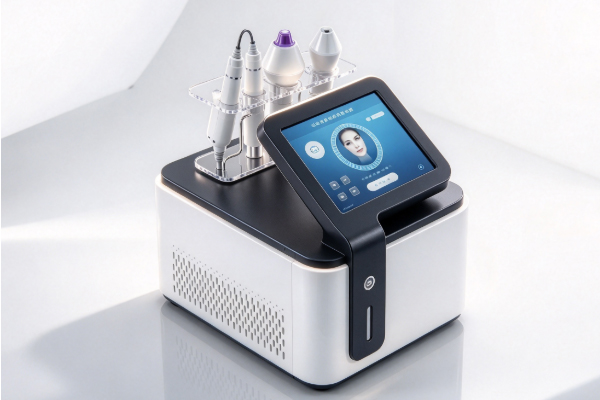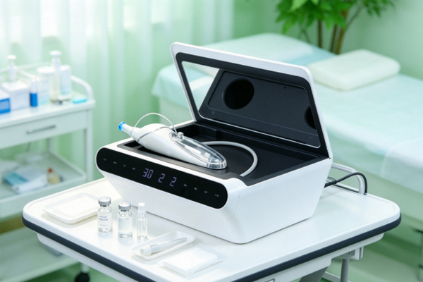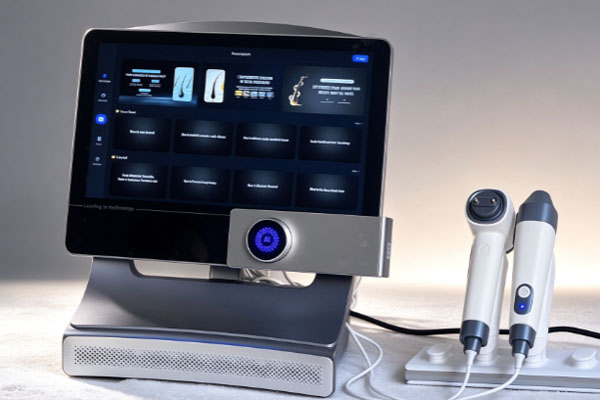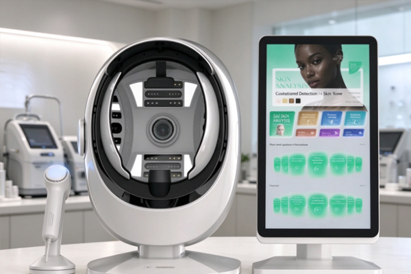
When examining skin with the naked eye, visible dark spots, enlarged pores, and redness are relatively easy to identify. However, some variations in skin tone, features around hair follicles, and subtle texture changes may be less noticeable. A skin analyzer uses imaging modes such as white light, ultraviolet (UV) light, or polarized light to capture the appearance of different facial areas, providing additional information for beauty consultations and skin condition management. Understanding what these devices can reveal helps beauty clinics tailor consultations and care recommendations to clients’ needs. However, results depend on device capabilities and imaging conditions. They provide supplementary visual information and cannot replace medical diagnosis.
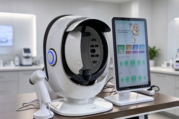
What Pigmentation Concerns Can a Skin Analyzer Observe?
Some pigmentation features are less visible under ordinary lighting. Different imaging modes can provide additional information to help observe pigmentation distribution across the face.
Subtle Differences in Pigmentation Distribution
UV imaging can help reveal certain pigmentation features that are difficult to distinguish with the naked eye. Comparing images of the forehead, cheeks, and nose can help identify differences in pigmentation distribution. Darker areas under UV light may indicate greater melanin absorption, but color alone cannot determine the type or depth of pigmentation. Saving images also allows skin appearance to be compared under similar conditions over time. These images can document pigmentation features but cannot independently determine the cause of dark spots or predict how they will develop.
Uneven Skin Tone and Pigmentation-Related Features
Uneven skin tone may be associated with pigmentation distribution, sun exposure, or post-inflammatory skin changes. A skin analyzer can help display some of these regional differences.
- Comparing facial areas: Compare skin tone across different areas to identify regions that may require further attention.
- Considering actual skin conditions: Interpret images alongside sun exposure habits, recent skincare routines, and visible skin characteristics.
- Interpreting results carefully: Images alone cannot accurately distinguish every pigmentation concern or determine the medical nature of a dark spot.
Combining imaging results with actual skin conditions helps make consultations more targeted and reduces the risk of overinterpreting a single measurement.
What Pore and Hair Follicle Features Can a Skin Analyzer Observe?
Some skin analyzers use white-light, polarized-light, or UV imaging to help examine pore distribution, skin texture, and specific fluorescence patterns.
Visible Pores and Uneven Skin Texture
Different imaging modes can capture certain surface details, providing useful references for observing pores and skin texture.
- Pore distribution: Examine the appearance of pores around the nose, forehead, and cheeks.
- Surface texture: Observe localized roughness, fine lines, and uneven skin surfaces.
- Regional comparison: Compare images of different facial areas to document variations in skin texture.
Images can reveal visible characteristics, but pore appearance alone cannot establish the underlying cause.
Fluorescence Features Around Hair Follicles
Some devices use UV light to stimulate fluorescence from certain substances, helping observe features around hair follicles.
- Localized fluorescence: Examine fluorescence patterns in areas such as the forehead and nose.
- Porphyrin-related features: Certain porphyrins may produce orange-red fluorescence, which can serve as supplementary visual information.
- Combined analysis: Interpret these features alongside other imaging results and the actual appearance of the skin.
Fluorescence does not directly indicate sebum levels and cannot independently diagnose acne or determine whether an infection is present.
What Signs of Redness and Skin Aging Can a Skin Analyzer Help Observe?
Devices equipped with appropriate imaging functions can record certain changes in skin color and texture, providing additional information for skin condition assessments.
Localized Redness and Color Changes
Polarized-light or multispectral imaging can help display skin color distribution and localized redness. Comparing color differences around the cheeks and sides of the nose, together with white-light and other imaging results, can help track changes in the same area. Saving images under similar conditions makes subsequent comparisons more meaningful. Red areas in an image do not establish a diagnosis, and persistent or unusual redness should be assessed by a qualified professional.
Fine Lines and Changes in Skin Surface Texture
A skin analyzer can record certain fine lines and surface texture features to help monitor visible changes in facial appearance.
- Fine-line observation: Examine the distribution of fine lines around the eyes, forehead, and other areas.
- Texture recording: Compare the smoothness and surface condition of localized skin areas.
- Change comparison: Save images taken at different times to observe changes in visible features.
Imaging results are useful for documenting appearance but cannot independently determine a person’s actual skin age or measure collagen content.
How to Interpret Skin Analyzer Results Correctly?
A skin analyzer provides images and supplementary analytical information. The usefulness of its results depends on device capabilities, imaging conditions, and how the findings are interpreted.
Selecting Imaging Modes According to the Observation Objective
Different light sources reveal different information. Select the imaging modes supported by the device according to the skin features being examined.
- White-light mode: Observe visible dark spots, fine lines, and overall skin tone.
- UV mode: Examine certain light-absorption differences and specific fluorescence patterns.
- Polarized-light mode: Depending on device capabilities, help observe skin texture, color variations, and some vascular-related features.
Combining appropriate imaging modes can provide more information than relying on a single image, but it cannot guarantee the detection of every hidden skin concern.
Standardizing Imaging and Monitoring Unusual Changes
Imaging conditions and substances on the skin’s surface may affect the results, making standardized procedures important.
- Maintain consistent imaging conditions: Keep facial positioning, lighting, and device settings as consistent as possible.
- Reduce external interference: Consider how cosmetics, sunscreen, and other surface residues may affect certain imaging results.
- Seek professional assessment: Consult a qualified medical professional about progressively worsening pigmentation, significant redness, or unusual skin lesions.
Standardized imaging and careful interpretation improve the usefulness of skin records and help prevent image features from being mistaken for medical diagnoses.
A skin analyzer can supplement visual skin assessments through different imaging modes, helping beauty clinics document pigmentation distribution, pore texture, localized redness, and certain fluorescence features. When selecting equipment, clinics should consider their business needs, available imaging modes, image clarity, and analytical functions while understanding the limitations of the results. Guangzhou Cosnext Technology Co., Ltd. provides information on skin analysis equipment to help beauty industry professionals explore device configurations and their potential applications.