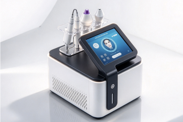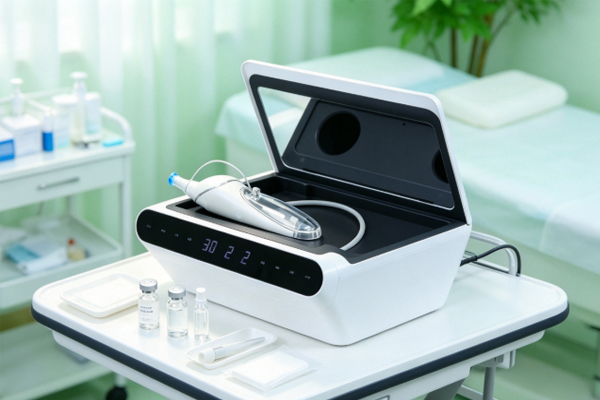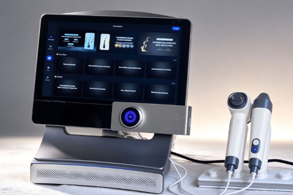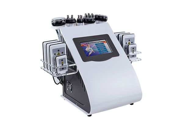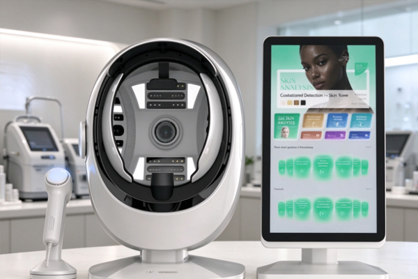
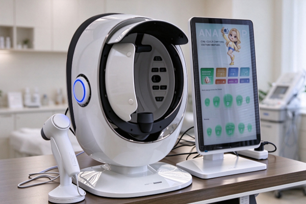
The same face can reveal noticeably different skin features under different lighting conditions. Standard photographs can capture visible spots, pores, and fine lines, but they may not fully reveal certain pigmentation patterns, localized color variations, or specific fluorescence features. Skin analyzers use imaging methods such as white light, ultraviolet light, and polarized light to capture facial images from different perspectives, providing more visual information for skin analysis. Understanding the role of each lighting mode helps beauty businesses select suitable analysis equipment, make client consultations more targeted, and explain results more clearly.
Why Do Different Lighting Conditions Reveal Different Skin Features?
When light interacts with the skin through reflection, absorption, and scattering, it produces different imaging information. Capturing facial images under multiple lighting conditions helps overcome the limitations of relying on a single light source.
White Light Records Visible Skin Conditions
White-light imaging closely resembles the way skin appears during everyday visual observation, making it suitable for documenting the overall condition of the face.
- Assess overall skin tone: Record facial brightness distribution, visible spots, and localized color differences.
- Capture surface details: Help examine the appearance of pores, fine lines, and skin texture.
- Create reference images: Provide a baseline for comparison with other lighting modes, making it easier to compare the appearance of the same area.
White-light images help beauty consultants understand a client’s visible skin condition, but they cannot reveal every subtle optical feature.
Ultraviolet and Polarized Light Provide Additional Imaging Information
Ultraviolet and polarized light use different imaging principles and are suited to observing different skin features. Ultraviolet imaging uses light absorption and fluorescence characteristics to help reveal pigmentation differences and fluorescence patterns. Parallel-polarized light preserves surface reflection information, highlighting skin texture and details. Cross-polarized light reduces specular reflection, helping reveal color features associated with pigmentation and blood vessels. These modes provide complementary information, but no single lighting method can reveal every structure beneath the skin’s surface.
What Is the Purpose of Capturing Images Under Multiple Lighting Conditions?
The value of multi-light imaging lies in recording skin features from different perspectives. This helps users develop a more comprehensive understanding of facial skin rather than relying on a single standard photograph.
Help Identify Subtle Pigmentation Differences
Some pigmentation patterns are less visible under ordinary lighting. Specific imaging modes can increase the visual contrast of certain areas.
- Compare different facial areas: Examine pigmentation features on the forehead, nose, and cheeks.
- Add supporting observations: Compare white-light and ultraviolet images to identify differences in certain pigmentation-related features.
- Track skin changes: Save images under similar shooting conditions to monitor changes in appearance.
Images provide supplementary information only. Color and brightness patterns alone cannot accurately determine the type, depth, or cause of pigmentation.
Record Texture, Redness, and Fluorescence Features
Multi-light imaging expands the range of observable features, allowing different aspects of the skin to be examined separately.
- Texture and pores: Use suitable visible-light or polarized-light modes to examine surface details.
- Localized redness: Use polarized-light and other imaging methods to help record facial color distribution.
- Specific fluorescence: Some devices can display fluorescence associated with substances such as porphyrins, providing additional clues for analysis.
This information helps organize observations of a client’s skin condition, but it cannot replace professional assessment or medical diagnosis.
How Can Images Captured Under Different Lighting Conditions Support Skin Analysis?
Capturing multiple images is not simply about increasing the number of photographs. More importantly, comparing different imaging modes helps reduce the limitations of relying on a single visual impression.
Improve the Completeness of Analysis Through Image Comparison
Features captured in the same facial area under different lighting conditions can complement one another, helping users examine skin characteristics more systematically.
- Use baseline images: Review the overall appearance under white light before referring to features shown in other modes.
- Separate observation priorities: Examine pigmentation, surface texture, and localized color changes individually.
- Avoid one-sided conclusions: Do not automatically interpret bright spots, dark areas, or red regions in a single image as definite skin problems.
Multiple images provide a broader basis for observation, but they do not replace professional interpretation.
Keep Records for Future Skin Condition Comparisons
Images captured using standardized procedures can serve as records over time, helping beauty businesses communicate changes in skin appearance to clients.
- Maintain consistent imaging conditions: Keep facial positioning, device settings, and lighting conditions as consistent as possible.
- Compare the same areas: Focus on identical facial regions and the same imaging modes when reviewing images taken at different times.
- Consider actual skincare practices: Review images alongside the client’s skincare records and observed skin condition.
Inconsistent shooting conditions may cause image differences, so not every change should be attributed to the skin itself.
What Should You Consider When Choosing a Multi-Light Skin Analyzer?
The number of lighting modes a device offers is not the only measure of its practical value. Imaging consistency, the purpose of each mode, and the interpretation of results also affect everyday performance.
Select Imaging Functions Based on Business Needs
When purchasing a skin analyzer, beauty businesses should evaluate its configuration according to their consultation priorities and operating procedures. Confirm that white-light images clearly show skin tone, spots, and texture. Check which features the additional ultraviolet and polarized-light modes are designed to observe. Assess whether image display, regional comparison, and record-keeping functions meet everyday consultation needs. Understanding the practical purpose of each feature is more useful than simply seeking a larger number of imaging modes.
Prioritize Standardized Operation and Accurate Interpretation
Proper operating procedures are essential for obtaining consistent images that can support skin assessment. Prepare the face and capture images according to the device’s instructions. Do not treat algorithmic markings, color distribution, or fluorescence images as diagnostic conclusions. For progressively worsening pigmentation, significant redness or swelling, or unusual skin lesions, clients should be advised to seek assessment from a qualified medical professional. Standardized device use improves the comparability of image records and helps clients understand the results more accurately.
Skin analyzers capture facial images under different lighting conditions to take advantage of the distinct imaging characteristics of each light source and reveal information that a single photograph may not fully show. This provides a more comprehensive reference for observing pigmentation, recording texture, and discussing skin conditions. For beauty businesses, choosing equipment with clear imaging, suitable functions, and standardized operating procedures can improve the consultation experience and strengthen service professionalism. Guangzhou Cosnext Technology Co., Ltd. provides information about skin analysis equipment to help beauty businesses understand device functions and their practical applications.