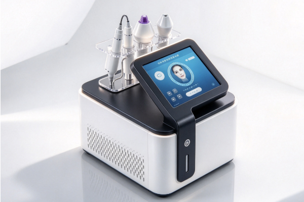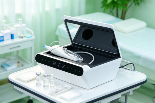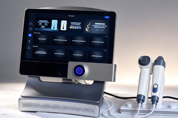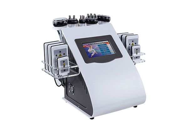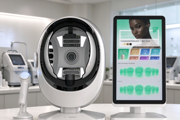
Facial pigmentation issues often appear as localized spots, uneven skin tone, or variations in color intensity. Visual observation alone may not fully capture subtle differences across facial areas or support consistent comparisons over time. A skin analyzer uses facial image capture, optical imaging, and image processing to help observe pigmentation-related features, providing a reference for skin assessment and skincare consultations. Understanding the device’s imaging methods, analysis results, and limitations helps beauty institutions standardize their assessment procedures, enables clients to better understand their skin’s appearance, and supports informed equipment selection.

How Does a Skin Analyzer Identify Facial Pigmentation Features?
A skin analyzer records facial information through light sources and imaging systems. Different optical modes can reveal different visual characteristics, and their actual performance depends on the device’s configuration.
Observing Skin Tone Under Standard and Polarized Light
Standard-light imaging displays visible skin tone, the appearance of pigmented spots, and their distribution. Some devices also feature polarized-light modes to reduce the impact of surface reflections on images.
- Standard-light imaging: Observes overall skin tone, localized spots, and color variations across the face.
- Cross-polarized imaging: Suppresses some specular reflections from the skin surface, helping visualize pigmentation- and redness-related features.
- Image comparison: Compares the visual characteristics of the same facial area under different imaging modes.
Polarized imaging can provide additional information beyond standard images, but it cannot independently determine the specific skin layer in which pigmentation is located.
Using Specific Wavelengths to Obtain Additional Information
Some devices use light sources at specific wavelengths and optical filters to help reveal certain features that may be difficult to observe under standard lighting.
- Specific ultraviolet imaging: Depending on the device design, it can reveal certain fluorescence or pigmentation-related features.
- Fluorescence image analysis: Displays fluorescence characteristics produced under specific conditions, with capabilities determined by the optical configuration.
- Multi-mode comparison: Combines information from different images to support a more comprehensive understanding of facial skin characteristics.
Different imaging methods have different applications. These images alone cannot establish a diagnosis of a pigmented skin disorder.
How Does a Skin Analyzer Assess Pigmentation Distribution?
After capturing images, the device can organize facial features through regional identification and color analysis, providing a reference for examining the distribution of pigmented spots.
Observing the Location and Shape of Pigmented Spots
Facial spots may be concentrated in specific areas or distributed across the face. Recording their visible characteristics helps support subsequent comparisons and assessments.
- Identifying key areas: Marks facial regions that require ongoing observation.
- Recording shape and appearance: Examines spot size, borders, and distribution patterns.
- Comparing localized colors: Compares spots with surrounding skin to identify differences in color intensity.
This information helps document the visible characteristics of facial pigmentation, but the cause of a spot cannot be determined from its appearance alone.
Understanding Pigmentation Intensity and Analysis Scores
Pigmentation may present differently across skin layers, but color intensity in an image does not directly indicate the actual depth of pigmentation. When interpreting regional markers, scores, or comparison results generated by a device, users should understand what each indicator means, the reference population used, and the applicable limitations. Skin tone, age, and image-capture conditions may also affect the results. A single score cannot establish the severity of a skin concern or replace professional diagnosis. Results should be interpreted by considering image features, device capabilities, and professional assessment together.
How to Track Pigmentation Changes with a Skin Analyzer
Ongoing records help users observe changes in facial appearance. To improve comparability between images captured at different times, image-capture conditions should be kept as consistent as possible.
Establishing a Standardized Baseline Image Record
During the initial assessment, images should be captured according to the device’s instructions, and relevant observations should be recorded.
- Maintaining consistent facial positioning: Keep the head angle, shooting distance, and positioning method as consistent as possible.
- Standardizing imaging settings: Control the light source and related parameters according to the device’s requirements.
- Reducing surface interference: Prepare the skin appropriately before imaging to prevent oil-related reflections or cosmetic residue from affecting the images.
Standardized records provide a more reliable reference for future comparisons.
Comparing Images Across Different Stages
During follow-up assessments, the same device and imaging modes should be used whenever possible, with comparisons focused on the same facial areas. Changes in facial positioning, lighting, surface reflections, and image-processing methods may affect how pigmented spots appear. The analysis should distinguish differences caused by capture conditions from actual changes in the skin. A change in image color alone is not sufficient to establish the effectiveness of a skincare treatment.
What Features Should You Consider When Choosing a Skin Analyzer?
When selecting equipment, beauty institutions should evaluate their assessment needs, operating procedures, and maintenance requirements rather than focusing solely on image clarity.
Understanding Imaging Configurations and Analysis Capabilities
Devices may differ in their light sources, filtering methods, and image-processing functions. Buyers should confirm the specific configuration before making a purchase.
- Checking imaging modes: Identify the optical imaging methods available and understand their intended uses.
- Reviewing analysis functions: Look for features such as regional marking, image storage, and comparisons across different assessment stages.
- Clarifying the scope of use: Understand which assessment tasks the results can support and what limitations apply.
Matching the device’s actual capabilities to the institution’s service offerings helps maximize its practical value.
Evaluating Operating Procedures and After-Sales Support
Ease of operation, maintenance requirements, and service support can all affect daily assessment workflows. Before purchasing, institutions can review equipment training, image-capture requirements, cleaning and maintenance procedures, and after-sales arrangements to ensure the features meet their operational needs. If a pigmented spot appears abnormal, continues to change, or requires further investigation, assessment by a qualified medical professional is recommended.
A skin analyzer can assist in observing facial pigmentation features, storing assessment images, and recording changes over time. However, results must be interpreted in light of the device’s capabilities and professional judgment. Choosing equipment with suitable functions, standardized operating procedures, and reliable maintenance support can help beauty institutions improve the quality of their skin analysis services. Guangzhou Cosnext Technology Co., Ltd. offers beauty equipment products and can support relevant institutions in understanding equipment configurations and usage requirements.