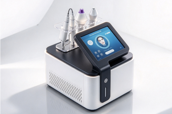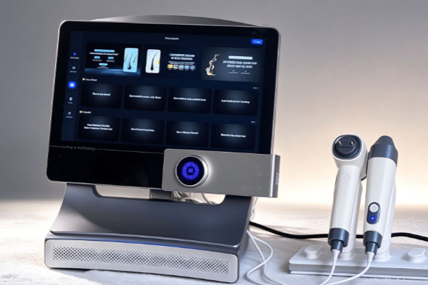
A hair analysis device is an examination tool used to observe the scalp surface, follicular openings, and hair characteristics. It is commonly found in beauty salons, scalp care centers, and professional hair management facilities. The device typically uses optical magnification, high-definition imaging, and image display technology to present scalp details that are difficult to see with the naked eye on a screen. This helps operators observe oil buildup around pores, keratin accumulation, hair distribution, and the condition of the scalp surface. In everyday use, scalp pores generally refer to the openings of hair follicles on the skin’s surface. Hair follicles are part of the skin structure, and hair grows from them. Sebum, shed keratinocytes, and other substances may also accumulate around follicular openings.
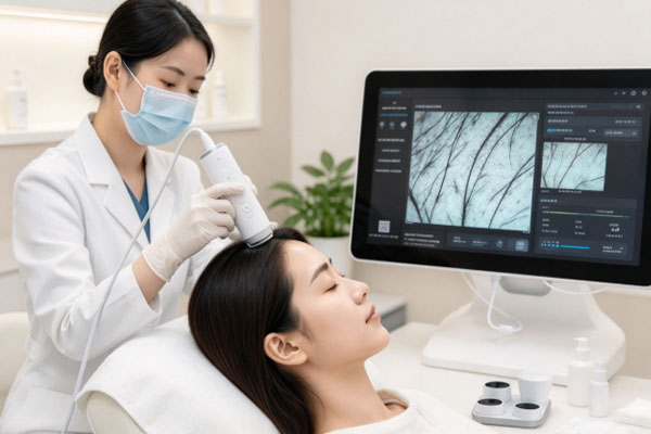
A hair analysis device can help observe these visible features, but images alone cannot directly determine the internal structure of hair follicles, their health, or future hair growth. When using a hair analysis device to examine scalp pores, operators generally need to select an appropriate examination area, adjust the probe position, magnification, and image clarity, and then compare images captured from different locations. A standardized examination procedure not only helps improve image recognition but also reduces assessment errors caused by hair obstruction, oil reflection, or improper probe positioning.
Basic Principles of Examining Scalp Pores with a Hair Analysis Device
Hair analysis devices primarily use optical magnification and image acquisition technology to examine the scalp surface. Some devices are also equipped with image annotation, data recording, and analysis software, which can help operators save examination results and compare images captured at different times.
Identifying the Locations of Hair Follicle Openings
In magnified scalp images, hair follicle openings usually appear where hair strands emerge from the skin, sometimes with slight depressions or visible outlines around them. Because scalp thickness, hair distribution, and sebum conditions vary across different areas, the appearance of follicular openings is not always the same. During examination, operators need to distinguish follicular openings from skin texture, dandruff, and substances attached near the hair roots. Not every circular or dot-shaped structure should be identified as a pore.
Observing Surface Features Around Pores
A hair analysis device can help operators observe whether there is visible oil buildup, keratin accumulation, redness, or other noticeable changes around follicular openings. This information helps document the condition of the scalp surface but cannot be used independently to diagnose skin diseases.
Opening contours: Observe whether follicular openings are clearly visible and whether obvious deposits are present around them.
Sebum distribution: Check for oily reflections or visible oily substances around the hair roots.
Keratin and dandruff: Observe whether loose or firmly attached white or pale yellow flakes are present on the scalp surface.
Surrounding skin: Check for noticeable redness, signs of irritation, or other abnormal appearances. Recommend further professional assessment when necessary.
Operating Procedure for Examining Scalp Pore Conditions with a Hair Analysis Device
Obtaining clear and useful scalp images requires not only good imaging performance but also appropriate selection of examination areas and standardized operation. Operators should adjust the probe and magnification according to the examination purpose and avoid assessing the condition of the entire scalp based on a single image.
Selecting Appropriate Scalp Examination Areas
Operators generally select areas such as the frontal hairline, crown, sides, or occipital region according to the examination requirements. Hair density, sebum distribution, and the appearance of follicular openings may vary between these areas, so the examination location should be selected according to the specific needs. Before examination, the hair can be gently parted to expose the scalp area being examined and prevent strands from obstructing the imaging field. If before-and-after comparisons are required, the same examination locations should be recorded whenever possible to facilitate the observation of image changes.
Adjusting Magnification and Image Clarity
After positioning the probe near the scalp, operators need to adjust the focus, light source, and magnification according to the device instructions so that follicular openings and surrounding skin texture appear clearly. If the magnification is too low, fine details may be difficult to identify. Excessive magnification, however, may narrow the field of view and make it difficult to assess the overall condition of the surrounding skin. Operators should also pay attention to probe stability and light reflection. Scalp oil may produce strong reflections that interfere with the identification of local contours. If images appear blurry or show excessive reflection, the shooting position and imaging conditions should be adjusted before examination continues.
Comparing Different Areas and Saving Records
After examining one area, operators can continue capturing images from other areas according to the examination purpose. Some devices support image storage, location annotation, or examination report generation for future reference. When comparing images captured at different times, it is important to keep the shooting position, magnification, and lighting conditions as consistent as possible. The time of the last hair wash and the use of scalp care products should also be recorded. This helps reduce differences caused by external conditions and prevents image changes from being directly interpreted as changes in follicular conditions.
Common Features to Observe in Scalp Pore Images
A hair analysis device provides magnified images of the scalp surface, but different visual features need to be interpreted in context. The clarity of follicular openings, the presence of oil or keratin deposits around them, and changes in local skin color may all be affected by cleansing habits, sebum production, imaging conditions, and the condition of the scalp.
Whether Hair Follicle Openings Are Clearly Visible
The visibility of follicular openings depends on hair positioning, substances attached to the scalp surface, and imaging conditions. Some openings may be covered by dandruff or oil, while others may not appear clearly because of improper focusing. Therefore, temporarily failing to see a clear opening in an image does not mean that the follicle has closed or lost its ability to produce hair. Operators should first rule out hair obstruction, blurry images, and surface residue, and recapture images when necessary.
Whether Oil and Keratin Deposits Are Present Around Pores
Sebum, dandruff, or keratin deposits may appear near follicular openings. A hair analysis device can help observe the appearance and distribution of these substances, but their exact composition generally cannot be determined from images alone. If the scalp frequently develops excessive dandruff, noticeable itching, redness, or pain, an assessment by a dermatologist should be considered. Simply removing visible deposits around pores does not necessarily address the underlying cause of these symptoms.
Frequently Asked Questions
Q1: Can a Hair Analysis Device See Inside Hair Follicles?
Answer: It can generally observe only the follicular openings and their surrounding surface features. Conventional optical imaging devices cannot fully display the deeper structures of hair follicles or directly measure their internal physiological conditions. If further evaluation of the follicles or a scalp condition is needed, a qualified medical professional should select an appropriate examination method based on the specific situation.
Q2: Do Larger-Looking Pores Indicate an Unhealthy Scalp?
Answer: Pore appearance alone is not enough to make this determination. The size and visibility of follicular openings are affected by individual differences, hair thickness, sebum deposits, and imaging magnification. A comprehensive assessment should also consider scalp color, dandruff, hair condition, and related symptoms.
Q3: Can a Hair Analysis Device Determine Whether Pores Are Clogged?
Answer: The device can help identify visible oil or keratin deposits near follicular openings, but images alone cannot confirm whether a hair follicle is genuinely clogged. If images show substantial local deposits, operators can record their appearance and interpret the findings alongside scalp cleansing habits and other visible signs.
Q4: Should You Wash Your Hair Before Examining Scalp Pores?
Answer: This depends on the examination purpose and the facility’s requirements. If the goal is to observe everyday sebum distribution, the examination facility may recommend avoiding hair washing for a specified period. If the goal is to examine the scalp after cleansing, a different preparation procedure may be used. Before the examination, confirm the specific requirements and inform the operator when you last washed your hair and which scalp care products you have recently used.
A hair analysis device helps examine follicular openings, surrounding oil, keratin deposits, and changes in the scalp surface through magnified imaging. A standardized procedure involves selecting examination areas, adjusting the probe, capturing images, and recording results, followed by comparisons between images from different locations. Clear imaging conditions and a relatively consistent examination procedure help improve the comparability of observations. When using these devices in beauty salons and scalp care centers, attention should be paid to probe cleanliness, image quality, and the objective interpretation of examination results.
Operators should not determine whether hair follicles are healthy or whether a hair loss disorder is present based solely on pore size, oiliness in one area, or a single image. A hair analysis device is better suited as an auxiliary tool for observing scalp appearance and recording routine care conditions. If persistent hair loss, noticeable redness and swelling, severe itching, or scalp pain occurs, a professional assessment by a dermatologist should be sought promptly. Combining device observations with symptom records and professional examinations provides a more reasonable understanding of the visible condition of scalp pores.