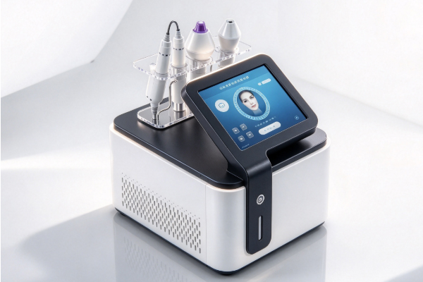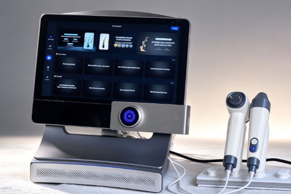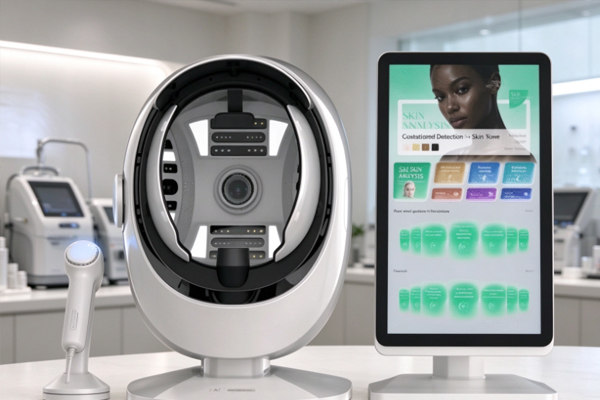
A hair analysis device is used to observe scalp conditions, hair distribution, and hair strand characteristics. It is commonly found in beauty salons, scalp care centers, and professional hair management facilities. These devices typically use high-definition cameras, optical magnification technology, or image analysis functions to capture localized images of the scalp and hair. They help operators observe hair follicle openings, scalp oil, keratin buildup, hair strand thickness, and hair density. Compared with visual inspection alone, a hair analysis device provides clearer close-up images that can serve as a reference for subsequent care recommendations.
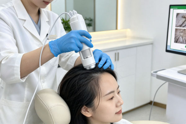
A hair analysis device typically takes a few minutes to more than ten minutes to examine hair. If the goal is simply to observe localized scalp and hair conditions, the process may take only 3–5 minutes. If multiple scalp areas need to be photographed, hair strand characteristics compared, and a report generated, the entire process usually takes 10–20 minutes. The exact duration depends on the device’s functions, the number of areas examined, hair cleanliness, and the operator’s level of experience. However, the time required for device photography and image collection is not exactly the same as the time needed for a complete hair assessment. A professional examination may also include basic information registration, an explanation of the results, and a discussion of care recommendations.
Examination Time for a Hair Analysis Device
The examination time of a hair analysis device generally depends on the purpose of the examination. Routine scalp observation and a more comprehensive hair assessment differ in their operating procedures and the time required.
Localized Scalp Examination: Approximately 3–5 Minutes
A localized examination is mainly used to quickly observe the scalp surface and the area around the hair roots. The operator selects areas such as the frontal hairline, crown, or other locations that require observation, then uses magnified images from the analyzer to examine oil distribution, keratin buildup, and the visible characteristics around hair follicles. This procedure is relatively simple and is suitable for an initial observation before treatment. It helps the operator understand the basic condition of the scalp. However, localized images only reflect the condition of the areas being examined and cannot directly represent the condition of the entire scalp.
Multi-Area Examination: Approximately 10–20 Minutes
If the crown, both sides of the scalp, forehead, and occipital region need to be examined, the operator generally needs to adjust the probe position for each area and complete image collection and comparative analysis. Some devices can also save images taken before and after treatment for subsequent comparison. Multi-area examinations provide more comprehensive observation records, but the time required still depends on the device’s capture speed, image analysis functions, and the examination items involved. These time estimates are intended as a reference for common procedures and are not a universal standard applicable to every device.
Main Steps in Hair Analysis Device Examinations
The examination process generally includes pre-examination preparation, probe imaging, image observation, and explanation of the results. Different brands vary in their operating interfaces, automatic analysis functions, and report formats, but the basic procedures are usually straightforward. Arranging the examination steps properly can reduce repeated photography and make the collected images easier to observe and record.
Preparation before testing
The operator will ask about the user’s scalp condition and main concerns, such as excess scalp oil, increased dandruff, thinning hair strands, or changes in hair volume. The operator then selects the areas to be examined according to the purpose of the examination and confirms whether the hair and scalp are suitable for image collection.
Preparation time: Usually approximately 1–3 minutes, although more complex situations may require additional time.
Preparation tasks: These include confirming the examination areas, parting the hair, and checking whether the device probe is clean.
Precautions: If a large amount of styling product remains on the scalp or certain scalp care products have been used recently, the user should inform the operator so that the visible features in the images can be interpreted appropriately.
Probe Imaging and Image Collection
After preparation, the operator places the examination probe over the areas to be observed and uses magnified imaging to examine details on the scalp surface and around the hair roots. Some devices can switch between different magnification levels to help observe both broader scalp conditions and localized hair strand characteristics. At this stage, the examination time mainly depends on the number of areas being photographed. If only one area needs to be examined, the process can usually be completed quickly. If multiple locations need to be recorded and images at different magnification levels saved, additional time will be required.
Image Analysis and Explanation of Results
After image capture, some hair analysis devices can use software to organize images, display relevant parameters, or generate examination reports. The operator explains the results based on the visible scalp characteristics and hair condition. It is important to understand that image analysis results serve as reference information for observation and assessment. Not all devices can accurately determine the specific causes of hair problems. For persistent hair loss, localized patchy hair loss, obvious redness or swelling, scalp pain, or similar symptoms, further evaluation by a dermatologist is recommended.
What Factors Can Change the Examination Time of a Hair Analysis Device?
The operating time of a hair analysis device is not fixed. In addition to the device’s imaging speed, the examination objectives, image quality, and level of detail provided when explaining the results can also affect the duration of the entire process. Some professional scalp assessment services may take approximately 20–45 minutes or even longer, but this time usually includes consultation, examination, and discussion of the results rather than device photography alone.
The complexity of the testing items
Basic observation: Mainly involves checking visible scalp oil, dandruff, and the condition around the hair roots. The procedure is relatively simple.
Comprehensive observation: Requires images from different areas and comparisons of hair density, strand thickness, and localized differences, so it takes relatively longer.
Image recording: If a personal examination record needs to be created, images saved, and current results compared with previous records, additional time must be allocated for data organization.
Device Functions and Operator Proficiency
Devices equipped with automatic image capture, data organization, and report generation functions can reduce some manual operating time. However, automatically generated parameters still need to be checked against image quality. The accuracy of examination conclusions should not be assumed solely based on software output. Operators who are familiar with probe positioning, capture distance, and image adjustment methods can also reduce repeated photography. For first-time users of the equipment, device setup and operation checks may require additional time.
Scalp Cleanliness and Examination Environment
Oil, flakes, sweat, and styling product residue on the scalp surface may affect the observation of certain image details. If the hair blocks the examination area, the operator may also need to part the hair again or adjust the probe position. There is no need to perform intensive cleansing or special treatments independently before the examination. Users can prepare their hair according to the examination facility’s instructions and explain their recent shampooing, coloring, perming, and scalp care routines in advance. If the examination is intended to assess persistent hair loss or scalp abnormalities, records of related symptoms and changes can also help professionals interpret the results.
Frequently Asked Questions
Q1: Can a Hair Analysis Device Complete an Examination in 5 Minutes?
Answer: Some basic examinations can be completed within this time. If only one or a few areas need to be observed, 5 minutes may be sufficient for image collection. However, if multi-area comparisons, parameter analysis, and report interpretation are also required, the process will usually take longer.
Q2: Can the Results Be Viewed Immediately After an Examination with a Hair Analysis Device?
Answer: Most devices with real-time imaging functions can display scalp images during the examination. If the device supports automatic report generation, some results may also be displayed shortly after the examination ends. However, image viewing, data interpretation, and professional assessment are separate steps. Immediate display should not be considered equivalent to a complete diagnosis.
Q3: Is It Necessary to Wash My Hair Before an Examination with a Hair Analysis Device?
Answer: Whether you need to wash your hair depends on the examination purpose and the facility’s requirements. If the goal is to observe the scalp’s natural oil distribution, the examiner may recommend not washing your hair on the day of the examination. If the goal is to observe the scalp after cleansing, a different preparation method may be used. Excessive cleansing or deliberately leaving large amounts of oil on the scalp may cause the examination results to differ from its usual condition.
Q4: How Much Time Should I Allow When Booking a Hair Analysis Device Examination?
Answer: It is recommended to allow 15–30 minutes, depending on the service provided. If the appointment includes a detailed consultation, comprehensive assessment, and written report, allowing 30–45 minutes is advisable. The exact duration depends on the examination facility’s procedures and the device’s functions. You can confirm whether an explanation of the results is included when making the appointment.
The time required for a hair analysis device to examine hair mainly depends on the examination scope and the services provided. Basic image collection may take only a few minutes, while multi-area observation and explanation of the results usually require around ten to twenty minutes. A comprehensive assessment that includes a detailed consultation may take half an hour or longer. Different examination purposes require a clear distinction between device operating time and the duration of the complete service, as the length of an examination alone does not determine the professionalism of the equipment. When selecting a hair analysis device, it is useful to consider image clarity, magnification range, image storage functions, data recording capabilities, and ease of operation.
Examiners should describe visible scalp and hair strand characteristics objectively and should not use a single image as the sole basis for diagnosing a disease. If the device is used for long-term scalp care records, images should be saved under similar lighting conditions, magnification levels, and scalp locations to facilitate subsequent comparisons. If noticeable hair loss, persistent itching, redness, swelling, or other abnormal symptoms occur, professional medical evaluation should be sought promptly. By allocating examination time appropriately, collecting images according to standardized procedures, and interpreting results correctly, a hair analysis device can better support routine observation and scalp care assessment.