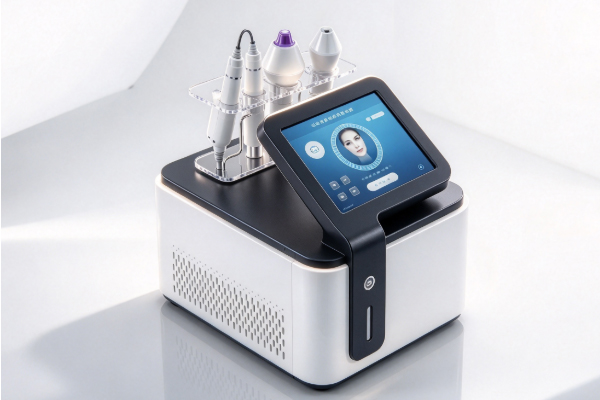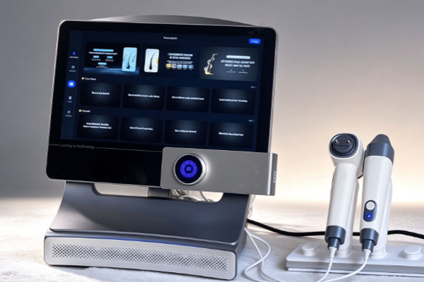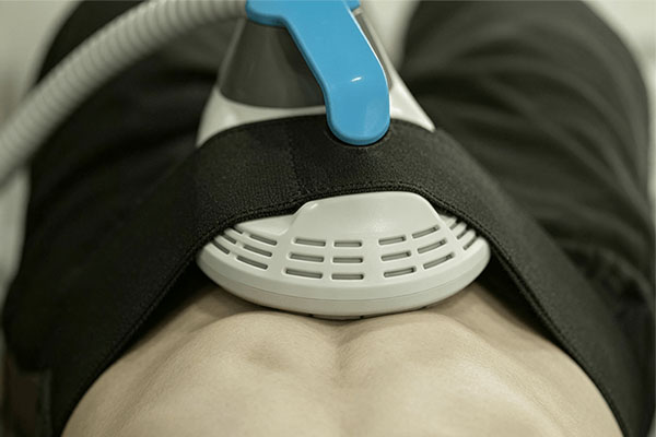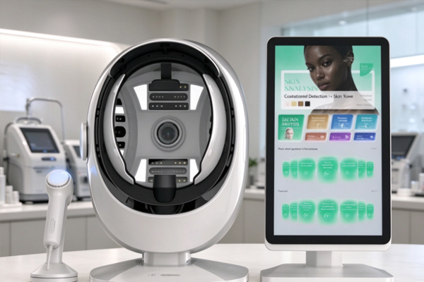
A hair analysis instrument is a professional testing device used to observe scalp conditions and hair characteristics. It is widely used in beauty care, scalp management, and hair care services. The device typically combines optical magnification, image acquisition, and digital image processing to magnify details of the scalp surface, areas around hair follicle openings, and individual hair strands. This helps care professionals understand scalp cleanliness, oil distribution, hair density, and hair condition. Compared with ordinary visual observation, a hair analysis instrument provides clearer images of specific areas, offering a visual reference for scalp condition assessment.
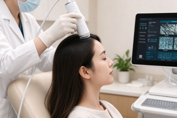
The working process of a hair analysis instrument usually includes image acquisition, image display, software analysis, and result recording. During an examination, the operator places the probe on the scalp area to be observed. The built-in camera and lighting system capture images, which are then transmitted to a display screen or supporting software. Some devices also use image recognition functions to assist in analyzing hair count, hair thickness, and localized scalp conditions. Different models vary in magnification, light source configuration, testing functions, and data analysis capabilities. Therefore, the specific examination items depend on the device configuration.
Core Working Principles of a Hair Analysis Instrument
A hair analysis instrument mainly relies on an optical imaging system to capture detailed information about the scalp and hair, then uses digital processing technology to display the collected images. The device magnifies details that are difficult to identify with the naked eye, allowing operators to examine the scalp surface and hair root areas more clearly.
Optical Magnification and Image Acquisition
Hair analysis instruments are usually equipped with macro lenses or microscopic imaging probes that allow the scalp to be observed at different magnification levels, depending on the device design. Some devices also feature LED light sources that provide stable illumination for the examination area, reducing the interference caused by changes in ambient lighting and helping maintain image clarity.
Optical Magnification: The lens magnifies details of the scalp surface and hair strands, making it easier to observe pore distribution, keratin buildup, and hair structure.
Image Acquisition: The camera converts the examination area into digital images, providing data for subsequent display and analysis.
Auxiliary Lighting: The built-in light source improves the brightness of the examination area, making scalp texture and hair details clearer.
The lenses, light sources, and magnification levels used by different devices are not exactly the same. Some basic models mainly provide magnified observation, while professional models may support multiple magnification levels, different lighting modes, and continuous image recording.
Digital Image Processing and Result Presentation
The captured images are transmitted to a display screen, computer, or supporting software. The software can enlarge, reduce, display specific areas of, and save images. Some models also offer image annotation, regional comparison, and automatic recognition functions. The main purpose of digital image processing is to make examination images easier to observe and record. For example, operators can compare hair distribution across different scalp areas or save images taken before and after treatment to help identify visible changes during the care process. Image processing does not directly provide a medical diagnosis. Image clarity, probe position, hair obstruction, and examination conditions can all affect the observation results. Standardized operation helps improve image usability and makes the examination results more useful as a reference.
Examination Procedures and Main Functions of a Hair Analysis Instrument
The process of obtaining images and generating examination results with a hair analysis instrument usually involves several basic steps. A standardized examination procedure not only helps produce clear images but also facilitates subsequent scalp care assessments.
Scalp Image Acquisition and Visual Observation
Before the examination begins, the operator usually selects a scalp area according to the examination purpose, such as the crown, hairline, or another area that requires observation. The probe is then placed on the target area, and the image is viewed on the display screen. The operator can adjust the probe position, focus, or lighting as needed. During the examination, the probe should remain as stable as possible, and the contact method and observation distance should follow the device instructions. If hair obstructs the examination area, it can be gently parted to expose the scalp surface. When comparing different areas, consistent image acquisition conditions should be maintained as much as possible to reduce differences caused by changes in lighting and camera position.
Analysis of Scalp and Hair Conditions
After image acquisition, the operator can examine relevant indicators according to the device’s functions. The available examination items vary by model, but commonly include the following:
Scalp Surface Condition: Observe visible features such as oil coverage, dandruff buildup, residual keratin, and scalp texture.
Hair Density and Distribution: Examine hair arrangement through localized images. Some devices can assist in counting the number of hairs within a designated area.
Hair Thickness and Shape: Examine hair shaft diameter, variations in thickness, and certain visible signs of surface damage.
Image Recording and Comparison: Save images from different times or scalp areas to help care professionals compare conditions.
These examination results can help identify the visible characteristics of the scalp and hair. However, the full physiological condition of hair follicles cannot be determined from a single image alone. If the assessment involves the severity of hair loss, the hair growth cycle, or specific scalp disorders, further professional examinations and medical evaluation are necessary.
The Value of Hair Analysis Instruments in Scalp Care
The main value of a hair analysis instrument lies in converting visible scalp and hair conditions into more intuitive visual records, helping care professionals observe, document, and communicate their findings. Standardized use of the device can reduce reliance on subjective descriptions alone and make scalp care procedures clearer.
Assisting in the Development of Scalp Care Plans
Scalp conditions vary from person to person, and care needs are not always the same. A hair analysis instrument helps care professionals examine oil distribution, flaking, and hair arrangement on the scalp surface. Combined with the user’s daily cleansing habits and care needs, these observations can support appropriate care recommendations. For example, if the images show noticeable oil buildup on the scalp, the operator can further investigate the user’s cleansing frequency and product use. If significant flaking is observed, it is necessary to consider whether symptoms such as itching or redness are present rather than immediately attributing the condition to a dry scalp.
Improving Examination Records and Service Communication
In beauty salons, scalp management centers, and professional care facilities, hair analysis instruments can also be used to establish client examination records. By saving images of the same scalp area, care professionals can show clients visible scalp conditions and compare the results during subsequent visits. Standardized records help care professionals explain their observations more clearly and allow clients to understand visible changes before and after treatment. However, changes in images do not necessarily mean that scalp health has improved. Evaluating care results still requires consideration of actual symptoms, examination conditions, and professional assessment.
Frequently Asked Questions
Can a Hair Analysis Instrument Directly Detect Hair Follicle Health?
A hair analysis instrument can observe visible structures around hair follicle openings and the distribution of hair within a localized area. However, ordinary optical devices cannot directly reveal all internal hair follicle structures or determine whether a follicle has the ability to produce normal hair growth based on images alone. For persistent hair loss, localized thinning, or other abnormal symptoms, further evaluation by a qualified medical professional is recommended.
Why Do Different Hair Analysis Instruments Produce Different Results?
Differences in lens magnification, imaging resolution, lighting methods, software algorithms, and operating procedures can all lead to variations in examination results. If the probe position, lighting, or hair-parting method differs between examinations, the resulting images may also appear different. Therefore, consistent image acquisition conditions should be maintained during repeated examinations, and the device should be operated according to its instructions.
Does a Hair Analysis Instrument Require Supporting Software?
This depends on the specific device design. Some models can display magnified images directly on a screen and are mainly used for on-site observation. Other models come with computer or mobile software that supports image storage, examination record management, localized annotation, and data comparison. When selecting a device, users should confirm that its software functions, data storage methods, and ease of operation meet their practical needs.
The working principles of a hair analysis instrument mainly involve optical magnification, image acquisition, digital image processing, and examination result recording. The device uses a camera probe to capture localized images of the scalp and hair, then presents observable details through a display system and supporting software. This helps care professionals understand scalp surface conditions, hair distribution, and hair strand characteristics. Some professional models also offer image recognition, density counting, and examination record management functions, providing a more intuitive reference for scalp care assessments.
When using a hair analysis instrument, it is necessary to select a suitable device according to the examination purpose and pay attention to probe position, lighting conditions, and standardized operating procedures. For beauty and scalp care facilities, imaging clarity, magnification levels, software functions, and data management capabilities are all important considerations when selecting equipment. It is important to understand that a hair analysis instrument is an auxiliary examination tool and cannot replace professional medical diagnosis. Combining device observations with the user’s scalp symptoms and daily care practices allows care plans to be developed more appropriately and helps improve the standardization of scalp examination and management.